GoF mutations - Tubulointersitial kidney disease
Autosomal dominant tubulointerstitial kidney disease (ADTKD) is a rare disease characterized by kidney tubule injury and interstitial fibrosis, invariably progressing to kidney failure. ADTKD is caused by GoF mutations in the UMOD gene encoding uromodulin (UMOD), an extracellular matrix protein secreted by kidney tubular cells. Most UMOD mutations are missense leading to retention of misfolded protein in the endoplasmic reticulum (ER) and formation of uromodulin aggregates damaging the cells. ADTKD is thus a paradigm of toxic GoF disease, as supported by the tissue damage triggered by UMOD aggregates causing endoplasmic reticulum (ER) stress in patients and mouse models. Conversely, the lack of UMOD is not toxic, as shown by the absence of any damage in Umod KO mice. Using phenotypic assays, we will screen for compounds that i) affect trafficking of mutant UMOD, ii) affect ER stress and UPR, coupled to the release of pro-fibrotic factors, and iii) reduce levels of UMOD production. Confirmed hits will be matched with their molecular targets and prioritized according to their links with UMOD synthesis. Identifying pathways affecting UMOD production may provide new starting points for small-molecule chemistry approaches. Confirmed hits from the natural product collection will be used to determine lead compounds for in vivo experiments in KI mice using established readouts. In parallel, we will test oligonucleotide-mediated inhibition of UMOD (WT, mutant, or both) or RNA-editing approaches as a therapeutic strategy for ADTKD.
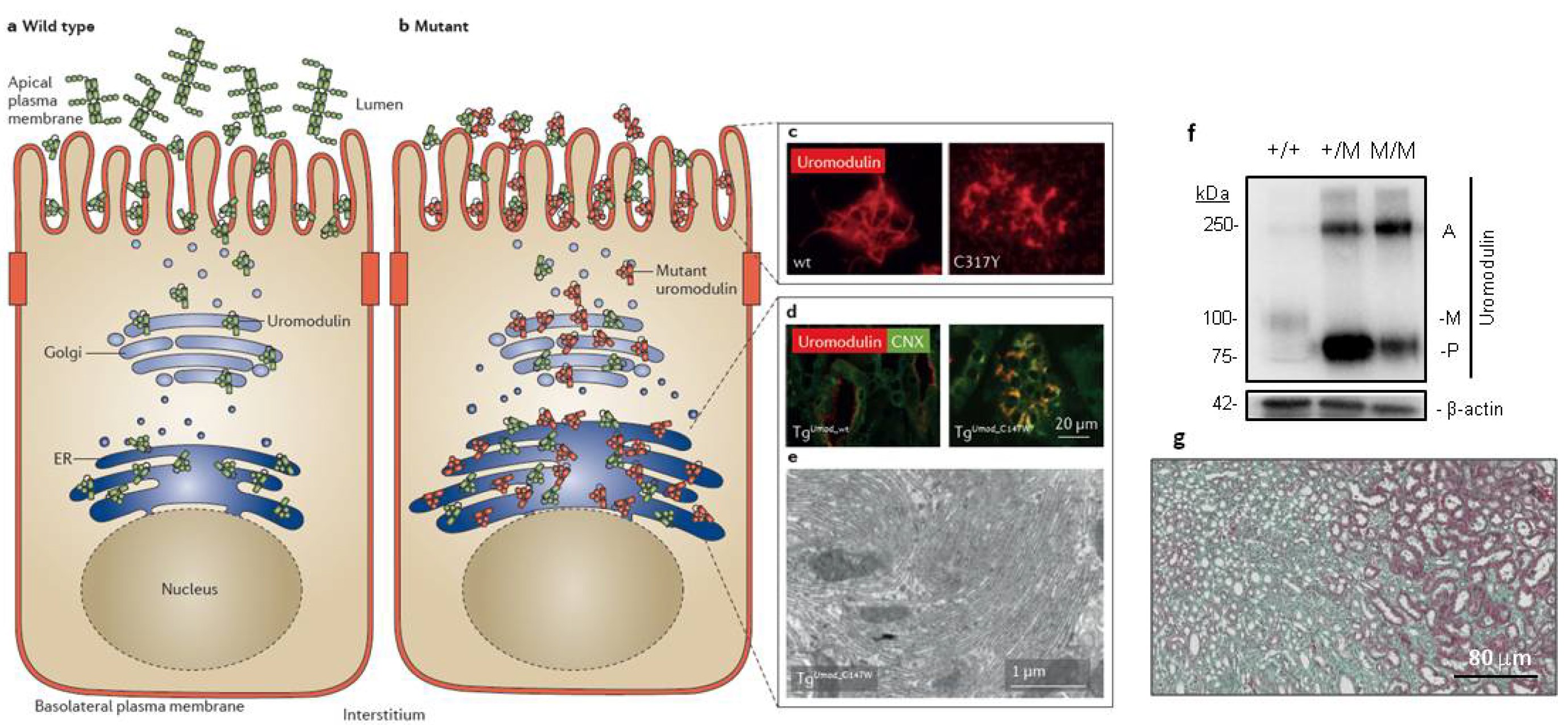
Figure 1. Pathophysiology of ADTKD due to mutations in UMOD. A) Normal trafficking of uromodulin in kidney tubular (TAL) cells, where its apical cleavage activates polymerization and formation of filaments in the urine. B) Mutations in UMOD causing ADTKD lead to ER retention and aggregation of mutant uromodulin, and lack of organized polymers in urine. C) immunofluorescence showing extracellular uromodulin secreted by MDCK cells stably expressing WT or mutant (C317Y) uromodulin isoforms. D) Immunofluorescence showing uromodulin in tubular segments of transgenic mice expressing WT (TgUmodwt) or mutant (TgUmodC147W) uromodulin. Colocalization with calnexin (CNX) indicates ER accumulation of mutant uromodulin, with ER hyperplasia shown by transmission electron microscopy (E). F) Immunoblotting for uromodulin in kidney extracts from WT (+/+) vs. mutant Umod (+/M, M/M) mice, showing changes in abundance of mature (M), precursor (P) and aggregate (A) forms of uromodulin. G) Trichrome staining evidencing kidney fibrosis in Umod+/M mice.